
Figure 5.
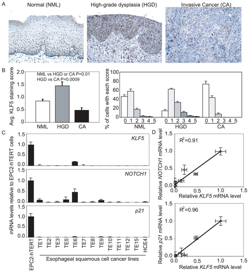
KLF5 is lost in human esophageal squamous cell cancer. (A) KLF5 was normally expressed in a gradient with highest levels in basal and suprabasal cells. In high-grade dysplasia, strong nuclear KLF5 expression was seen throughout esophageal epithelia; expression was lost in invasive esophageal squamous cell cancers. Magnification=100×. (B) Quantitatively, the average score for KLF5 staining per cell increased by 70% from normal (NML, n=6) epithelia to high-grade dysplasia (HGD, n=5), but decreased by 45% and 70%, respectively from NML or HGD to invasive squamous cell cancer (CA, n=6). Furthermore, 37% of NML, 11% of HGD, and 59% of CA cells had no staining, while the percentage with scores of 2 or greater was 17% for NML, 39% for HGD, and 6% for CA. (C) Esophageal squamous cell cancer lines lost KLF5, NOTCH1, and p21Waf1/Cip1 expression by quantitative real-time PCR. (D) KLF5 expression in esophageal squamous cell cancer lines was highly correlated with NOTCH1 and p21Waf1/Cip1 expression.