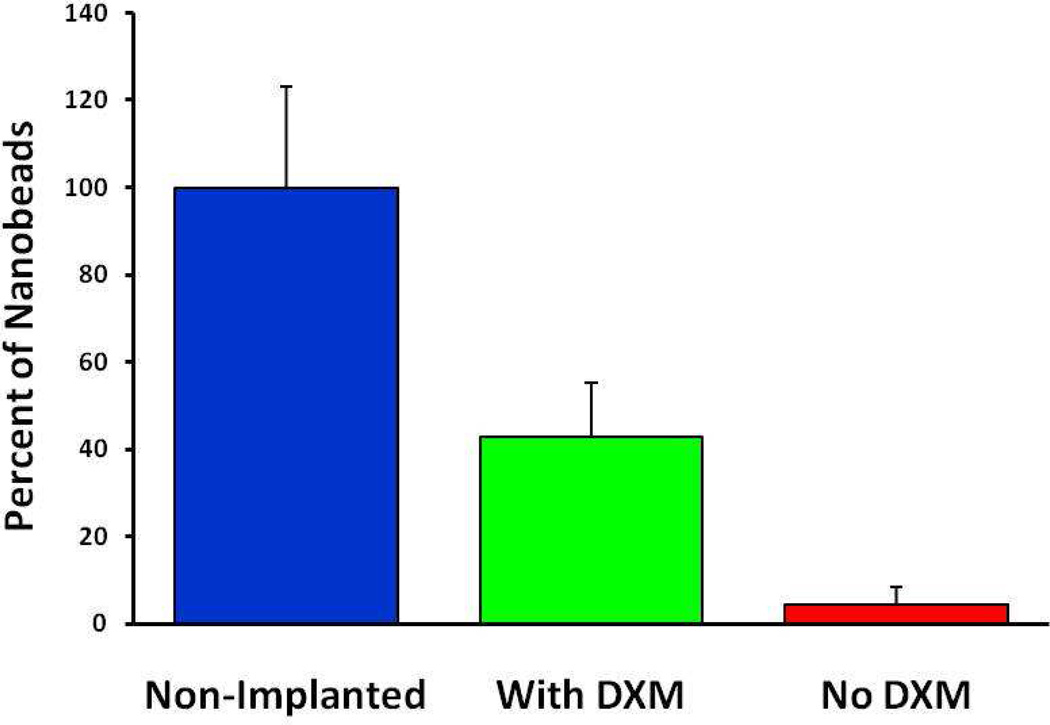
Fig. 6.
The number of fluorescent pixels in blood vessel images from non-implanted control tissues (blue) and implanted tissues perfused with (green) and without (red) dexamethasone: the data are normalized with respect to the average pixel count from the non-implanted controls. Tissue with and without dexamethasone were significantly different from one another and from non-Implanted tissue (ANOVA and Tukey posthoc test: F(2,12) = 57.1; p<0.00001).

