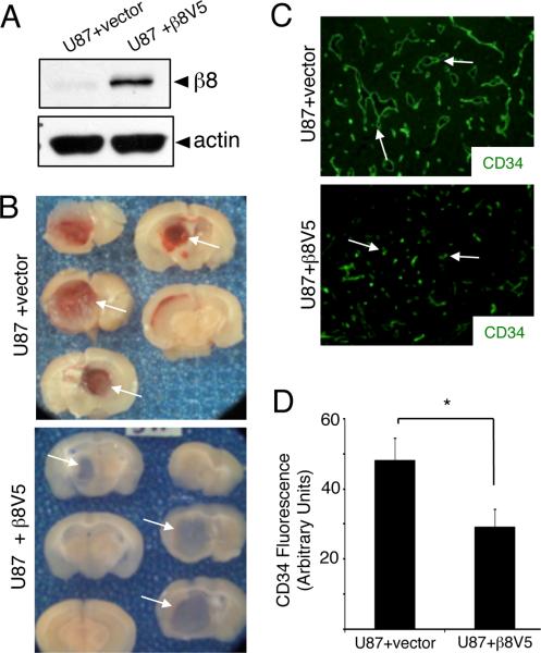
Figure 2. β8 integrin suppresses microvascular pathologies and hemorrhage in GBM.
(A); U87 cells were stably transfected with empty plasmid or a plasmid containing a cDNA encoding for a β8-V5 fusion protein. Detergent-soluble lysates were then immunoblotted with antibodies directed against anti-β8 integrin or anti-actin antibodies. Note the increased expression of β8 integrin protein. (B); U87 cells stably transfected with empty plasmid (top panels) or plasmid harboring a β8-V5 cDNA (bottom panels) were stereotactically injected into the striatum of immunocompromised mice (n=5 mice per cell type). Shown are gross images of slices from representative brains harboring tumors generated from each cell type. Note that the intratumoral hemorrhage evident in the U87 tumors is not evident in tumors overexpressing β8-V5 protein. (C); U87 tumors stably transfected with empty plasmid or plasmid expressing β8-V5 were immunostained with an anti-CD34 antibody to visualize vascular endothelial cells (n=3 different brains per cell type). Note the increased numbers of blood vessels and abnormal morphologies in U87 tumors transfected with empty plasmid versus the capillary-like morphologies in U87 tumors overexpressing β8 integrin. (D); Immunofluorescence intensities were quantified based on anti-CD34 antibody staining (shown at 200x magnification). Note the significant reduction of fluorescent intensity in U87 tumors forcibly expressing β8-V5 integrin protein (*p<0.0001). Error bars represent standard deviations.

