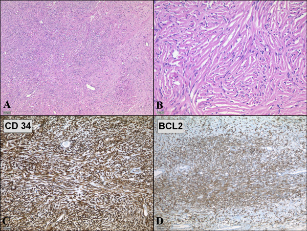
Figure 3.
Microscopic examination of solitary fibrous tumor of the pleura. (A, B) Microscopic specimen of the tumor shows solid proliferation of spindle-shaped fibroblastic cells in a patternless pattern. (Hematoxylin and eosin; magnification 40× and 200×) (C, D) Spindle-shaped tumor cells show strong positivities for immunohistochemical staining with CD34 (C) and BCL2 (D).

