Abstract
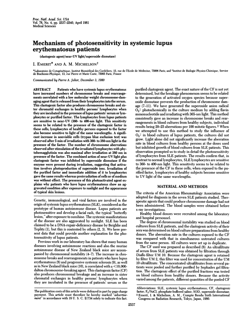
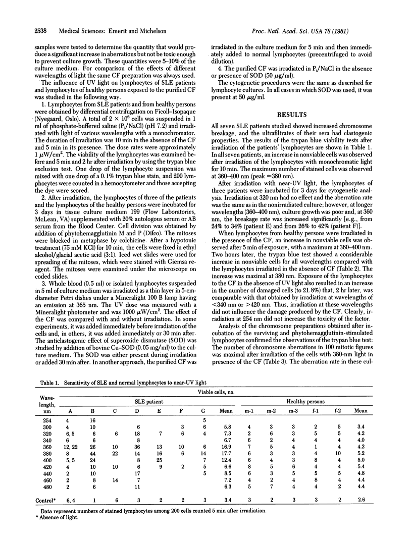
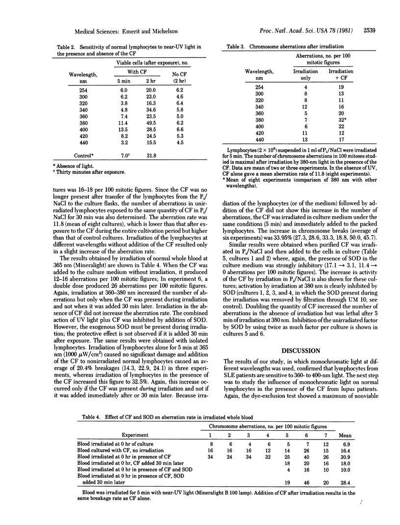
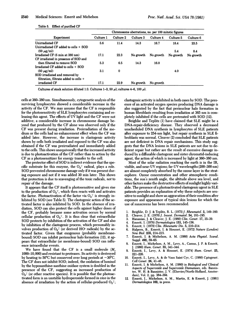
Patients who have systemic lupus erythematosus have increased numbers of chromosome breaks and rearrangements correlated with a low molecular weight chromosome-damaging agent that is released from their lymphocytes into the serum. This clastogenic factor also produces chromosome breaks and sister chromatid exchanges in healthy persons' lymphocytes when they are incubated in the presence of lupus patients' serum or lymphocytes or purified factor. The lymphocytes from lupus patients are sensitive to near-UV (360- to 400-nm light. This sensitivity seems to be related to the presence of the clastogenic factor in these cells; lymphocytes of healthy persons exposed to the factor also become sensitive to light of the same wavelengths. A significant increase in nonviable cells (trypan blue exclusion test) was observed after 5 min of irradiation with 360- and 380-nm light in the presence of the factor. The number of chromosome aberrations observed after stimulation of the irradiated lymphocytes with phytohemagglutinin was also maximal after irradiation at 380 nm in presence of the factor. The combined action of near-UV light plus clastogenic factor was inhibited by superoxide dismutase if the enzyme were present during irradiation, suggesting that activation involves photoproduction of superoxide ions. Irradiation of the purified factor and immediate addition of it to lymphocytes gave the same results whereas preirradiation of cells or of medium was without effect. The presence of this photoactivated agent explains why patients who have lupus erythematosus show an aggravated condition after exposure to sunlight and the appearance of typical skin lesions.
Full text
PDF


Selected References
These references are in PubMed. This may not be the complete list of references from this article.
- Beighlie D. J., Teplitz R. L. Repair of UV damaged DNA in systemic lupus erythematosus. J Rheumatol. 1975 Jun;2(2):149–160. [PubMed] [Google Scholar]
- Cleaver J. E. DNA damage and repair in light-sensitive human skin disease. J Invest Dermatol. 1970 Mar;54(3):181–195. doi: 10.1111/1523-1747.ep12280225. [DOI] [PubMed] [Google Scholar]
- Emerit I. Chromosomal breakage in systemic sclerosis and related disorders. Dermatologica. 1976;153(3):145–156. doi: 10.1159/000251109. [DOI] [PubMed] [Google Scholar]
- Emerit I., Levy A., Housset E. Breakage factor in systemic sclerosis and protector effect of L-cysteine. Humangenetik. 1974;25(3):221–226. doi: 10.1007/BF00281430. [DOI] [PubMed] [Google Scholar]
- Emerit I., Levy A., de Vaux Saint Cyr C. Chromosome damaging agent of low molecular weight in the serum of New Zealand black mice. Cytogenet Cell Genet. 1980;26(1):41–48. doi: 10.1159/000131420. [DOI] [PubMed] [Google Scholar]
- Emerit I., Michelson A. M. Chromosome instability in human and murine autoimmune disease: anticlastogenic effect of superoxide dismutase. Acta Physiol Scand Suppl. 1980;492:59–65. [PubMed] [Google Scholar]
- Halpern B., Emerit I., Housset E., Feinglod J. Possible autoimmune processes related to chromosomal abnormalities (breakage) in NZB mice. Nat New Biol. 1972 Feb 16;235(59):214–215. doi: 10.1038/newbio235214a0. [DOI] [PubMed] [Google Scholar]
- Hananian J., Cleaver J. E. Xeroderma pigmentosum exhibiting neurological disorders and systemic lupus erythematosus. Clin Genet. 1980 Jan;17(1):39–45. doi: 10.1111/j.1399-0004.1980.tb00112.x. [DOI] [PubMed] [Google Scholar]


