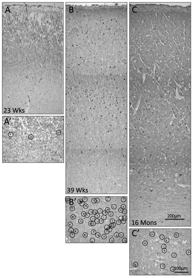
Figure 7.
Developmental changes in γ-aminobutyric acid (GABA)ergic neuronal density in the frontal association cortex. (A–C) Twenty-three weeks (mid-gestation) (A), 37 weeks (term) (B), and 16 postnatal momths (C), are shown. The density is markedly increased at term compared to midgestation or postnatally. A′–C′ are high power of A–C at Layer IV; each circle outlines a GABAergic neuron demonstrated by glutamic acid decarboxylase (GAD65/67) immunostaining.

