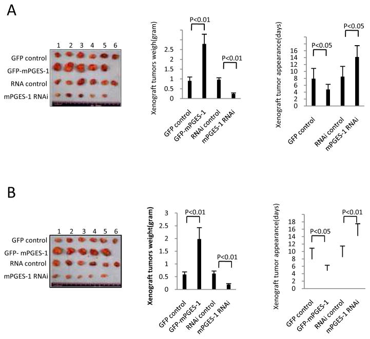
Figure 3. mPGES-1 promotes HCC growth in SCID mice.
Hep3B and Huh7 cells stably transfected with mPGES-1 expression or siRNA vectors were injected subcutaneously at armpit of SCID mice (100 μl cell suspension at a concentration of 1 × 10⁸ cells per ml in PBS). The mice were monitored for tumor formation (5 or 6 mice for each group) and the tumors were recovered 4 weeks after inoculation (A - Hep3B tumor; B - Huh7 tumor). The wet weight of each tumor and tumor appearance time (days) were determined for each mouse. The photographs of the transplanted tumors are shown at the left panels. The average weight of the xenograft tumors are shown at the mid panels (the data represent mean ± SEM, n = 5–6). The average onset time (days) of the xenograft tumors (days) are shown at the right panels (the data represent mean ± SEM, n = 5–6).

