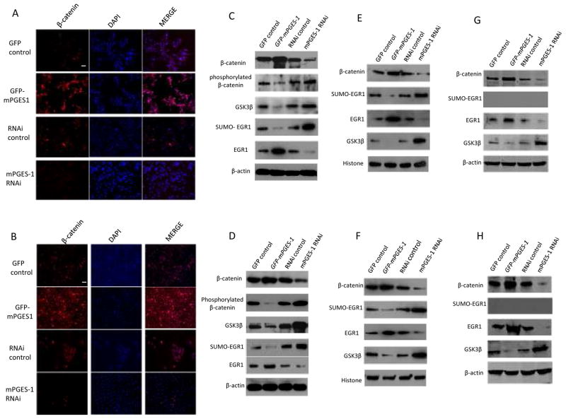
Figure 4. mPGES-1 increases β-catenin and EGR1 in human HCC cells.
A & B. Confocal microscopy showing immunofluorescence staining of β-catenin in Hep3B (A) and Huh7 (B) stable cell lines (with TRITC staining and DAPI counterstaining; original magnification×200; scale bar 10μm). The level of β-catenin was increased in mPGES-1 overexpressed cells but decreased in mPGES-1 knockdown cells.
C & D. Western blotting using whole cellular proteins from Hep3B (C) and Huh7 (D) cells stably transfected with mPGES-1 expression vector or RNAi vector. mPGES-1 overexpression increases β-catenin with concurrent reduction of GSK-3β and phosphorylated β-catenin. In contrast, mPGES-1 knockdown reduces β-catenin with concurrent increase of GSK-3β and phosphorylated β-catenin. Furthermore, mPGES-1 overexpression also increases EGR1 with simultaneous reduction of SUMO-EGR1, whereas mPGES-1 knockdown reduces EGR1 but increases SUMO-EGR1.
E & F. Western blotting using nuclear proteins from Hep3B (E) and Huh7 (F) cells stably transfected with mPGES-1 expression vector or RNAi vector. mPGES-1 overexpressed cells show increased nuclear levels of β-catenin and EGR1 but decreased nuclear levels of GSK-3β and SUMO-EGR1. An opposite pattern was seen in cells with mPGES-1 depletion.
G & H. Western blotting using cytoplasmic proteins from Hep3B (G) and Huh7 (H) cells stably transfected with mPGES-1 expression vector or RNAi vector. mPGES-1 overexpression increases β-catenin with concurrent reduction of GSK-3β. In contrast, mPGES-1 knockdown reduces β-catenin with concurrent increase of GSK-3β. Furthermore, mPGES-1 overexpression also increases EGR1, whereas mPGES-1 knockdown reduces EGR1. SUMO-EGR1 was not detected in cytoplasmic protein.

