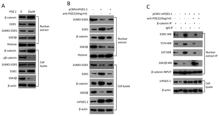
Figure 5. The effect of PGE2 in HCC cells.
A. Western blotting using whole cell lysates or nuclear extracts from Huh7 cells treated with 10μM PGE2 (Cayman Chemical Company) for 24 hours or the vehicle control (DMSO).
B. Huh7 cell line transfected with mPGES-1 expression vector were treated with 10ng/ml anti-PGE2 (highly specific for PGE2, purchased from Abcam, San Francisco, CA) or the vehicle control (DMSO) for 24 hours. The whole cell lysates or nuclear extracts were obtained for western blotting analysis.
C. Huh7 cell line transfected with mPGES-1 expression vector were treated with 10 ng/ml anti-PGE2 antibody or vehicle (DMSO) for 24 hours. The whole cell lysates or nuclear extracts were obtained for western blotting analysis. The nuclear extracts were subjected to immunoprecipitation with anti-β-catenin antibody followed by western blotting analysis for EGR1, TCF4, LEF1, and GSK-3β. Nuclear β-catenin was used as input control for immunoprecipitation; β-actin was used as internal control for routine western blotting. IP – immunoprecipitation; WB – western blotting.

