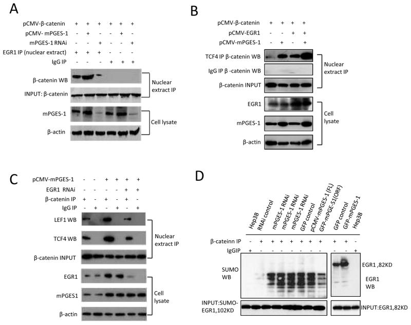
Figure 6. mPGES-1 enhances nuclear β-catenin association with EGR1, TCF4 or LEF1 in Hep3B cells.
Hep3B cells were transfected with different vectors (mPGES-1 expression or RNAi vector, β-catenin expression vector, EGR1 expression or RNAi vector, and control vectors) as indicated on the top of the panels. The nuclear extracts were obtained and subjected to immunoprecipitation by using specific antibodies or IgG as the control, followed by western blotting using specific antibodies as indicated for each panel. The nuclear extracts or whole cell lysates were also processed for routine western blotting analysis. IP – immunoprecipitation; WB – western blotting.
A. mPGES-1 increased the association between β-catenin and EGR1. Nuclear β-catenin was used as input control for immunoprecipitation.
B. mPGES-1 increases the interaction between β-catenin and TCF4 in the nuclei isolated from Hep3B stable cell lines; the interaction is enhanced by EGR1 overexpression. Nuclear β-catenin was used as input control for immunoprecipitation.
C. mPGES-1 increases the interaction between β-catenin and TCF4 or LEF1 in the nuclei isolated from Hep3B stable cell lines; the interaction is decreased in cells with EGR1 depletion. Nuclear β-catenin was used as input control for immunoprecipitation.
D. mPGES-1 increases β-catenin binding to EGR1, but not SUMO-EGR1. Western blots for SUMO-EGR1 (102 KD) or EGR1 (82 KD) were used as input controls.

