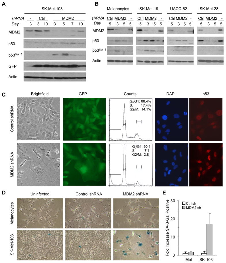
FIGURE 3. MDM2 genetic inactivation triggers melanoma growth arrest and senescence.
A,B, Protein immunoblots of the indicated cell lines that were either uninfected (−) or infected for GFP-lentiviruses coding for Control shRNA (Ctrl) or MDM2 shRNA. C, (Left panels) Representative brightfield and corresponding GFP fluorescent microphotographs (day 7) of SK-Mel-103 transduced with the indicated lentiviral vectors. (Middle panels) Cell cycle distribution as determined by flow cytometry (day 3). (Right panels) Visualization of p53 induction by immunofluorescence. D, SA-β-Gal staining in MDM2 shRNA-expressing SK-Mel-103 cells as compared to uninfected and Control shRNA cells. E, Corresponding quantification of cells exhibiting positive blue staining for SA-β-Gal. Data correspond to the mean ± SEM of cells from three experiments.

