
Fig. 5. Aβ peptide species conformation.
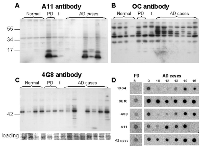
Western blots labeled with conformation-selective antibodies in same normal (n=4) and AD cases (n=7) used for Fig. 3. (A) Prefibrillar oligomers labeled with A11; (B) fibrillar oligomers labeled with the antibody OC. (C) Western blot for the N-terminal Aβ antibody 4G8 with Coomasie blue-stained membrane as loading control. (D) Dot blots using detergent-free sonicated synaptosome-enriched fractions; cases shown correspond to the lane number in (A) and (C), i.e., the PD case is the right-most case in lane 6 of Western blots.