
Figure 1.
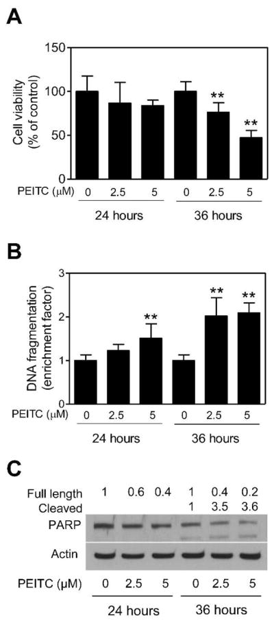
The PEITC treatment decreased viability of BRI-JM04 cells. (A) Viability of BRI-JM04 cells after 24- or 36-hour treatment with DMSO (control) or the indicated concentrations of PEITC as determined by trypan blue dye exclusion assay. Percent cell viability relative to DMSO-treated control is shown for each treatment. (B) Cytoplasmic histone-associated DNA fragment release into the cytosol after 24- or 36-hour treatment of BRI-JM04 cells with DMSO (control) or the indicated concentrations of PEITC. Results are expressed as enrichment factor relative to DMSO-treated control. (C) Western blotting for PARP cleavage using lysates from BRI-JM04 cells after 24- or 36-hour treatment with DMSO (control) or the indicated concentrations of PEITC. Numbers above bands represent changes in levels of full length and cleaved PARP relative to DMSO-treated control at each time point. Quantitative experiments (panels A and B) were done twice in triplicate and combined data from both experiments are presented as mean ± SD (n = 6). **Significantly different (P<0.01) compared with corresponding control by one-way ANOVA with Dunnett’s adjustment.