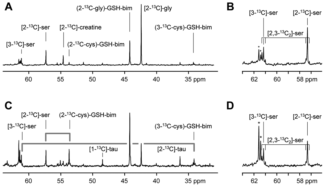
Figure 4.
High resolution 13C magnetic resonance spectra acquired from monobromobimane treated acid extracts of tumor (A, B) and liver (C, D) tissue samples taken from rats infused with [2-13C]-glycine at 1 mmol/kg/h for 24 h. The appearance of the label in the 2-carbon of the glycinyl-residue of glutathione-bimane (2-13C-gly)-GSH-bim, the 2-carbon of the cysteinyl-residue ((2-13C-cys)-GSH-bim) and the 3-carbon of the cysteinyl-residue ((3-13C-cys)-GSH-bim) are detected. The fate of the serine 13C-labels as they are metabolized through the transsulfuration pathway and appear in the cysteinyl-carbons of glutathione are indicated by the grey lines. Additional resonances are visible from taurine (tau). An expansion of the region around the serine peaks is shown (B,D). These show the presence of singly labeled [3-13C]-serine and [2-13C]-serine and smaller amounts of [2,3-13C2]-serine. The asterisks indicate peaks due to natural abundance glucose in the extracts.

