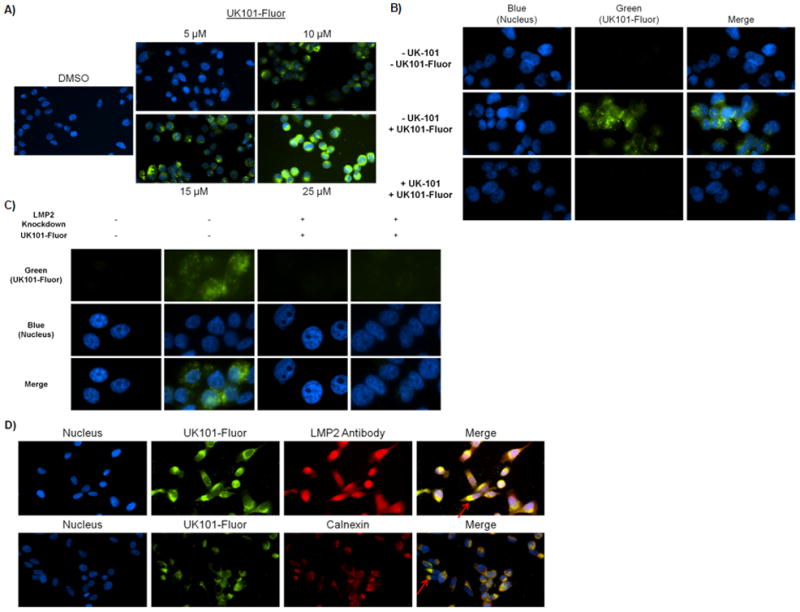
Figure 5.

UK101-Fluor (9) selectively binds to LMP2 in PC-3 cells. A) UK101-Fluor binding with LMP2 is dose-dependent. After extensive washing to remove unbound probe, binding was examined via fluorescence microscopy. Fluorescence intensity of all images was normalized to DMSO control. Of note, LMP2 binding was also observed in cells treated with 5 μM UK101-Fluor with a longer exposure time. B) Pre-incubation with UK-101 competes away the signal observed following treatment with UK101-Fluor alone. PC-3 cells pretreated with UK-101 or DMSO were subseqently treated with UK101-Fluor, fixed, and examined via fluorescence microscopy. Fluorescence intensity in all images was normalized to control (-UK-101/-UK101-Fluor). C) LMP2 knockdown results in decreased binding of UK101-Fluor. PC-3 cells transfected with either the empty pLKO vector (LMP2 Knockdown −) or the pLKO vector containing a short hairpin RNA against LMP2 (LMP2 knockdown +) were treated with DMSO or UK101-Fluor, fixed, and examined via fluorescence microscopy. Fluorescence intensity in all images was normalized to controls (UK101-Fluor −). D) UK101-Fluor cellular localization. Cells were incubated with UK101-Fluor and subsequently stained with antibodies recognizing LMP2 or calnexin, an endoplasmic reticulum (ER) marker. The cells were then fixed and examined via fluorescence microscopy. The arrows in the merged images point to areas of overlap between green and red staining, which gives a yellow color.
