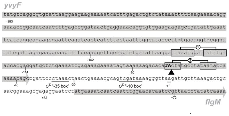
Figure 4. The PflgM promoter region.
The sequence is of the 3′ end of the yvyF gene and the 5′ end of the flgM gene from B. subtilis strain 3610 (gray arrows behind text). Underlined sequences indicate the predicted “−35” and “−10” promoter elements of the σD consensus sequence and the +1 transcriptional start predicted for the flgM gene in Fig S2. Boxes indicate inverted repeat sequences protected by DegU-P in the DNase footprint assay in Fig. 5B. Inverted triangle and bolded, capitalized sequence indicates the location of the TnΩ2723 transposon insertion that phenocopies a degU mutation and restores flagellin expression to cells mutated for SwrA and SwrB. Dashed lines indicate important positions relative to the electrophoretic mobility shift experiments in Fig. 5A.

