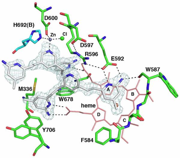
Figure 3.
The nNOS active site with one molecule of 3j bound above the heme and the other in the pterin binding pocket. The sigmaA-weighted Fo-Fc omit density map for 3j is shown at a 3.0 σ contour level. The ligation bonds around the new Zn2+ site and hydrogen bonds are depicted with dashed lines. Two alternate side chain conformations are shown for residue Tyr706. NOS dimerizes through the heme domains with the pterin binding in a pocket at the dimer interface. Residues in subunit A are depicted with green bonds and those of subunit B with cyan bonds. Four pyrrole rings of heme are labeled.

