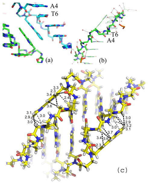
Figure 4.
(a) Pair of π-stacked bipyridine ligands in the crystal structure of Bipy PNA. The view of the backbone of Bipy PNA shows the orientation of backbone carbonyl of T6 towards the N-end of the PNA and the minor groove of the duplex in contrast to that of the carbonyl groups in A4 and G7, which are oriented towards the C end and away from the duplex. (b) PNA backbone for the central base pairs in Bipy PNA and water molecules (red crosses) that are situated closest to the PNA backbone; (c) Relative orientation of bipyridines with respect to the Bipy PNA duplex from which they are extruded.

