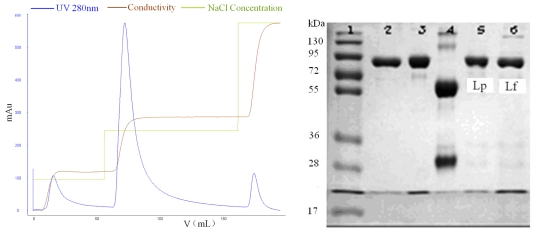
Figure 5.
Isolation of LF and Lp in a stepwise manner from the defatted colostrum using SPEC 70 SLS column (Φ 1.6 × 6 cm) after static adsorption and the analysis graph obtained from Quantity One software. SDS-PAGE: Lane 1, standard protein markers; Lane 2, Lf; Lane 3, Lp; Lane 4, IgG; Lanes 5 and 6, the isolates of first and second peaks eluted by different concentrations of NaCl.

