
Figure 1.
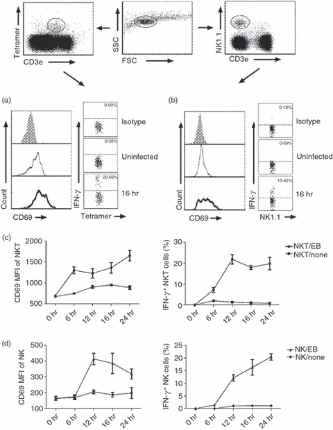
Natural killer (NK) and NKT cells were activated and produced interferon-γ (IFN-γ) following in vitro infection with Chlamydia muridorum. Ex vivo splenocytes isolated from naive C57BL/6 mice (three mice) were infected with live C. muridorum elementary bodies at a multiplicity of infection of 3 and cultured for 6–24 hr in complete culture medium. Control cells were cultured but not infected. Cells were analysed by multi-colour staining and flow cytometry at designated time-points. The different cell populations were gated as shown in dot plot and analysed for CD69 and intracellular IFN-γ. (a) Expression of activation marker CD69 on iNKT (PBS-57 glycolipid loaded CD1d tetramer+ CD3e+) cells and intracellular IFN-γ staining at 16 hr culture. Grey histograms show isotype control antibody staining, thin line shows uninfected control cells and thick line shows infected cells. (b) CD69 expression and IFN-γ production by NK (NK1.1+ CD3e−) cells at 16 hr culture. The mean fluorescence intensity (MFI) of CD69 expression and the percentage of IFN-γ production by iNKT cells (c) and NK cells (d) at different time-points after infection are represented. Data are shown as means of triplicate analysis ± SD and are representative of three independent experiments.