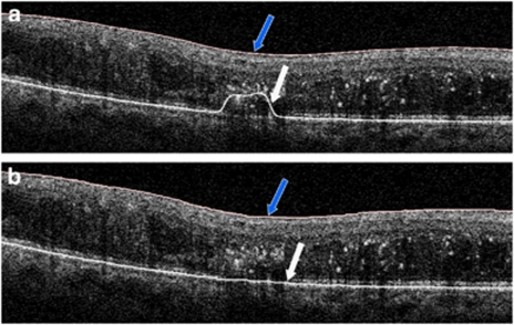
Figure 1.
(a) Uncorrected optical coherence tomography (OCT) B-scan with retinal segmentation boundaries generated by the Cirrus OCT instrument software. (b) Corrected OCT B-scan with manual placement of retinal boundaries; blue arrow indicates the inner limiting membrane boundary and the white arrow indicates the retinal pigment epithelial boundary. The color reproduction of this figure is available at the Eye Journal online.

