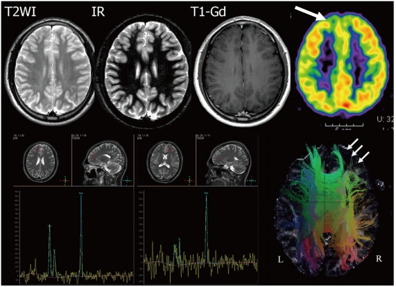
Fig. 12.
8-year-old male with intractable seizure. Conventional spin-echo and inversion recovery (IR) MRI shows no definite abnormality of frontal cortex. PET demonstrates decreased metabolism at right frontal cortex (thick arrow). MR spectroscopy describes increased choline level in right frontal cortex. Tractography of both hemispheres reveals decreased subcortical fiber connectivity in left frontal cortex (thin arrows). In this case, tractography was more sensitive than other conventional MRI modalities and it can be compared with PET or MR spectroscopy.

