Fig. 3.
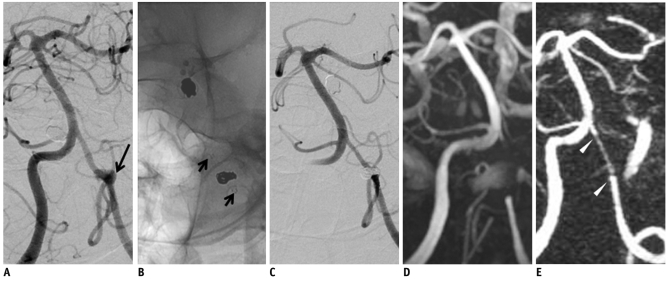
59-year-old male with aneurysm at V4 segment of vertebral artery.
A. Digital subtraction angiography shows broad-neck aneurysm (long arrow) at V4 segment of left vertebral artery involving posterior inferior cerebral artery origin. B. Stent-assisted coil embolization is successfully performed with Enterprise stent (short arrows). C. Digital subtraction angiography with 6-month interval shows patent stented segment and complete occlusion of treated aneurysm in left vertebral artery. D. 3D time of flight demonstrates complete signal loss causing non-visualization of stented segment of left vertebral artery. E. 4D MR angiography shows mild signal loss of stented segment and diffuse artifactual luminal narrowing (arrowheads). There is no evidence of recanalization of treated aneurysm.

