Fig. 4.
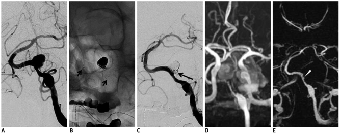
60-year-old male with large thrombosed aneurysm at vertebral artery.
A. Digital subtraction angiography shows only non-thrombosed portion of large aneurysm. B. Stent-assisted coil embolization is performed using Neuroform stent (short arrows). C. Digital subtraction angiography after coil embolization shows minimal residual neck filling (long arrow). D. 3D time of flight after 1-day interval demonstrates diffuse narrowing of stented segment of left vertebral artery. However, it is difficult to evaluate completeness of coil embolization due to T1 shortening artifact of thrombosed aneurysm. E. 4D MR angiography clearly visualizes stented segment of left vertebral artery. Residual neck of aneurysm (arrowhead) is also suspected.

