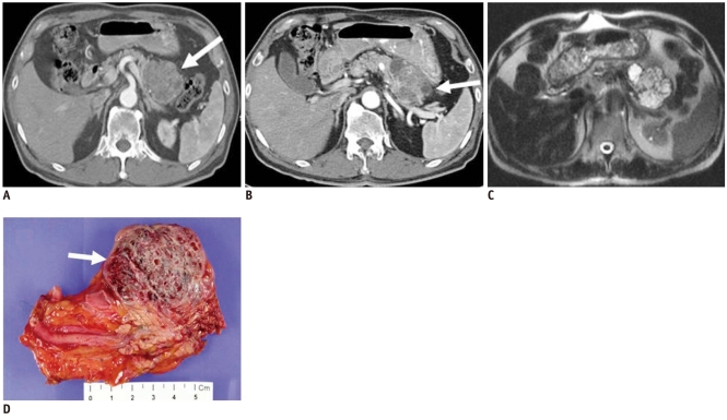
Fig. 10.
66-year-old man with microcystic serous cystadenoma manifesting as microcystic lesion.
A. Contrast-enhanced transverse CT during arterial phase shows hypervascular mass (white arrow) in tail of pancreas. It appears as solid lesion on CT, owing to innumerable cysts and fine septa. B. Note cystic portion in periphery of tumor (white arrow). C. Axial T2-weighted image reveals microcystic mass. D. Gross specimen shows multilocular, well-defined bulging mass containing multiple tiny cystic spaces and sanguineous clear fluid (white arrow), which are confirmed as microcystic serous cystadenoma.

