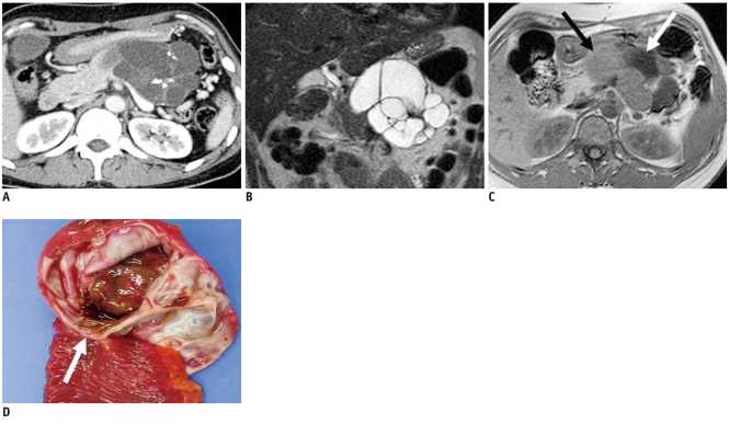
Fig. 11.
37-year-old woman with mucinous cystic neoplasm: pitfall in differentiating multilocular cystic lesions.
A. Contrast-enhanced CT image during venous phase shows multilocular lobulated cyst with internal septation and calcification in tail of pancreas. B. Coronal T2-weighted image reveals multilocular cystic mass. C. T1-weighted gradient-echo image shows multiple locules with different signal intensity (black and white arrows). D. Gross specimen shows multilocular cystic tumor with inner smooth and glistening surface (white arrow) which was confirmed as mucinous cystadenoma at microscopy.

