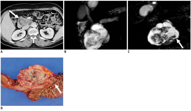
Fig. 12.
59-year-old-woman with branch duct type intraductal papillary mucinous neoplasm manifesting as mixed cystic and solid lesion.
A. Contrast-enhanced CT image during venous phase shows cystic mass and internal solid portion in head of pancreas (white arrow). B. Two-dimensional thick slab MR cholangiopancreatography image shows cystic mass in head of pancreas. Communication between cystic mass and pancreatic duct is not clear. C. Source image of 3D-MR cholangiopancreatography shows narrow tumor neck suggesting communication between tumor and main pancreatic duct more clearly than 2D-MR cholangiopancreatography (white arrow). D. Gross specimen shows whitish polypoid friable mass containing translucent mucoid materials. Tumor communicates with main pancreatic duct (white arrow) and was confirmed as branch duct type intraductal papillary mucinous neoplasm.

