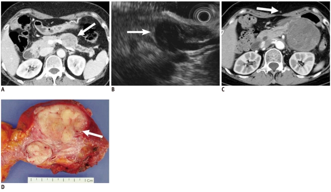
Fig. 2.
58-year-old woman with metastatic leiomyosarcoma manifesting as solid lesion.
A. Contrast-enhanced CT image during venous phase shows well-marginated, low attenuating solid tumor in pancreas (white arrow). B. Endoscopic US image depicts heterogeneously hypoechoic solid tumor (white arrow). Woman had multiple painless metastatic tumors in her stomach, chest, thigh and pancreas, which were too numerous to excise. Consequently, surgery was ruled out, and only chemotherapy was performed. C. Woman began to feel sick and mass increased in size at 1-year follow-up CT. Note metastatic nodule in subcutaneous layer (white arrow). D. Tumor was finally excised and appeared as whitish solid tumor in gross specimen and was confirmed as metastatic leiomyosarcoma (white arrow).

