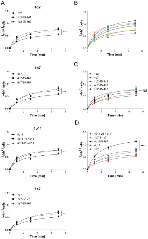
Figure 4. Bivalency enhances internalization.
(A–D) Internalization assay of selected VHHs. 1 µM VHH was incubated on cells at 37°C for 1, 3 or 7 min. Externally bound VHH was removed with 25 µg/ml trypsin for 45 min on ice. Internalized VHH was immunolabelled with an anti-myc tag and D-α-M-IR800 antibodies. Quantification of fluorescence is described in experimental procedures. Fluorescence intensities of the internalized amount is divided over the amount of cells (To-pro3 nuclear stain) and plotted against time. The signal of the irrelevant anti-hTfR VHH was deducted from the specific VHH signal and therefore represents the x-axis (n = 12) (s.e.m.'s are shown for all points).

