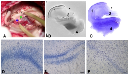
Figure 2. Tissue sampling and histopathological results.
In A, surgical view of the hippocampus: (1) body of the hippocampus/CA1; (2) head of the hippocampus; (3) dentate gyrus/fimbria (green dot); (4) parahippocampal gyrus (blue dot). B, MRI of surgically resected hippocampus; C– F Nissl-stained hippocampal slices. In B and C the location of tissue resection for genomic analysis is marked with an asterisk showing the resection in the CA3–CA4 transition. D–F Cytoarchitectural alterations of the granule cell layer in sclerotic hippocampi. D: cell dispersion. E: bilamination. F: cell loss. Calibration bar = 100 microns.

