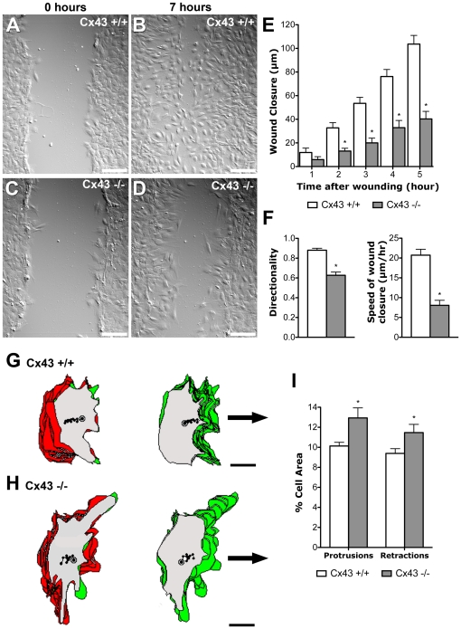
Figure 1. Wound closure assay shows defect in polarized cell movement in Cx43 KO MEFs.
(A–F). A wound scratch was introduced in confluent monolayers of Cx43 KO (n = 7 scratches) (C) or wildtype MEFs (n = 7 scratches) (A). After 7 hrs, wildtype MEFs have migrated into the gap to close the wound (B), while there are still extensive gaps in the wound edge of KO MEFs (D). Tracking the position of the advancing wound edge revealed a significant decrease in the rate of wound closure in the KO MEFs (E). There was also a marked decrease in the directionality and speed of wound closure (F). Asterisks indicate p<0.05 when comparing wildtype vs. KO MEFs. (G–I). Tracings of individual cells at the wound edge showed a distinct polarized cell morphology associated with wildtype cells (n = 42 cells) (G), with cytoplasmic protrusions (green) concentrated at the leading edge of the cell, facing the wound edge, while retractions of cell processes (red) were situated mostly at the ipsilateral or trailing edge of the cell. In contrast, in KO MEFs (n = 42 cells) (H), cytoplasmic protrusions and retractions were observed around the entire cell circumference, with extensive overlap between regions of protrusions and retractions, thereby indicating a defect in polarized or directional cell movement. Quantitative analysis showed this was associated with an increase in both cytoplasmic protrusions and retractions (I). Data presented as mean ± SEM. Scale bars in (A–D) represent 100 µm. Scale bars in (G, H) represent 10 µm.

