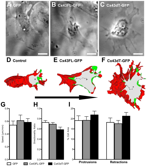
Figure 6. Neural crest cells expressing Cx43dT-GFP also show defects in polarized cell migration.
Neural crest cell explants from E8.5 wildtype mouse embryos were transfected with either Cx43FL-GFP or Cx43dT-GFP plasmid constructs and neural crest cell migration behavior was examined 24 hours after transfection. Motion analysis with the tracing of individual cells at the explants edge showed a distinct polarized cell morphology in GFP construct-transfected control cells (n = 11 cells) (A, D) or Cx43FL-GFP transfected cells (n = 12 cells) (B, E), with cytoplasmic protrusions (green) seen at the cell's leading edge, and retracting cell processes (red) in the cell's trailing edge at the ipsilateral side. In contrast, in cells expressing the Cx43dT-GFP construct (n = 21 cells) (C, F), cell protrusions and retractions were not as distinctly polarized. This defect in cell polarity in neural crest cells expressing the Cx43dT-GFP construct was associated with an overall increase in cell protrusive activity (I), a significant decrease in cell directionality (H) (i.e. a more randomized migration pathway), and a not significantly altered migration speed (G). Scale bars = 20 µm. Data presented as mean ± SEM.

