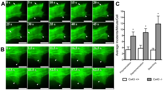
Figure 7. TIRF imaging show increased microtubule instability in Cx43 KO MEFs.
Time lapse TIRF imaging of Cx43 KO (A) and wildtype MEFs (B) transfected with a tubulin-GFP plasmid construct (see Movie S1 and Movie S2) showed increased microtubule polymerization (arrowhead in A), depolymerization (arrow in A), and searching events (multiple polymerization/depolymerization; arrowhead in B) in the Cx43 KO MEF. Quantification of the data obtained from the TIRF imaging is shown in (C) (Cx43 +/+ n = 13 cells, Cx43 −/− n = 22 cells). Data presented as mean ± SEM. Scale bars represent 5 µm.

