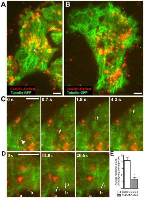
Figure 8. Two colour TIRF imaging show reduction in microtubule targeting to cell surface localized Cx43dT-DsRed.
NIH3T3 cells transfected with GFP-tubulin and either Cx43dT-DsRed or Cx43FL-DsRed were examined by two colour TIRF imaging. In Cx43FL-DsRed expressing cells, microtubules were centrally organized around a single MTOC (asterisk in A), while in cells expressing Cx43dT-DsRed, microtubules appear disorganized in distribution (B). Time-lapse TIRF imaging shows Cx43FL-DsRed (arrowhead in C) being transported to the cell membrane along microtubules (arrows in C) (see Movie S3). Also observed is microtubule polymerization and targeting (labeled 1-3 in D) to Cx43FL-DsRed plaques in the cell membrane (labeled a, b in D) (see Movie S4). Such microtubule targeting events were significantly decreased in NIH3T3 cells expressing Cx43dT-DsRed (n = 18) when compared with those expressing Cx43FL-DsRed (n = 16) (E). Data presented as mean ± SEM. All scale bars represent 5 µm.

