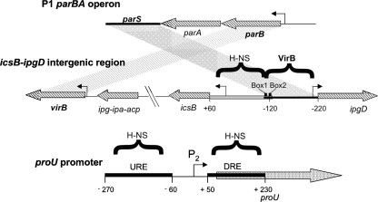
Fig. 1.
Comparison of the parBA operon from bacteriophage P1, the icsB-ipgD intergenic region from S. flexneri, and the proU promoter from E. coli. The parS centromere-like site is located downstream of the parBA operon in bacteriophage P1 (top). The P1 parS region corresponds to the VirB binding region in the S. flexneri virulence plasmid, and the P1 parB gene corresponds to the S. flexneri virB gene (middle) (3). The gray parallelogram denotes DNA sequence homology between parS and the S. flexneri VirB binding site; the striped parallelogram illustrates homology between the parB and virB genes. DNA sequences bound by the H-NS protein or by the VirB protein are indicated by horizontal brackets. The positions of the essential box 1/box 2 VirB binding and nucleation sites are shown. The regulatory region of proU is illustrated (bottom), together with the locations of the proU P2 promoter and the URE and DRE H-NS binding sites (gray boxes). The angled arrows represent promoters; the diagram is not drawn to scale.

