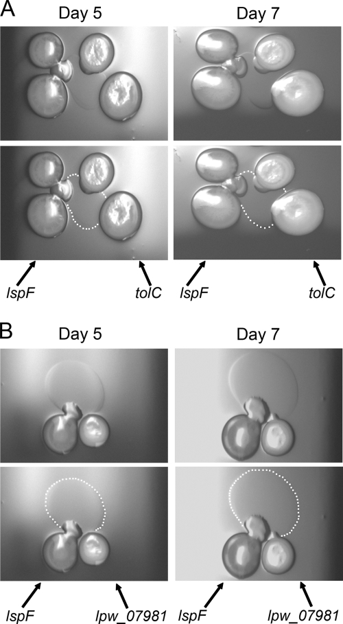
Fig. 5.
Surfactant secretion by an L. pneumophila lspF mutant when grown in the presence of an L. pneumophila tolC or lpw_07981 mutant. (A) As indicated by the arrows, two inocula of the lspF mutant NU275 and two inocula of the tolC mutant NU390 were spotted next to each other on a BCYE plate containing 0.5% agar. After incubation at 30°C for 5 days or 7 days, bacterial growth and surface translocation were imaged (top row). To help with visualization of the surfactant that was produced by the sliding lspF mutant, white dots have been added to mark the boundary of the spreading film (bottom row). The surfactant film was also evident when single spots of the two mutants were placed next to each other (data not shown). (B) As indicated, lspF mutant NU275 and lpw_07981 mutant NU402 were spotted next to each other on low-agar BCYE plates, and then after incubation at 30°C for 5 or 7 days, bacterial growth, surface translocation, and surfactant production were imaged. As in panel A, the bottom images depict the boundary of the film produced by the lspF mutant with white dots. The results depicted in panels A and B are representative of those obtained from three independent experiments.

