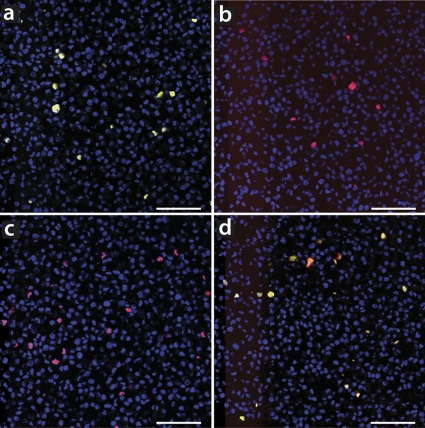
Fig. 6.
Specificities of genotypic oligonucleotides scrutinized on recombinant PCV2a- or recombinant PCV2b-infected cell layers in laser confocal microscopy. Shown are selected images from TMA block sections of formalin-fixed and paraffin-embedded genotype-specific transfected PK15 cell layers. PCV2a or PCV2b genotype-infected tissues were either stained with a combination of oligonucleotides AB (red) and A (green) as single colors (a and b) or separately with oligonucleotides AB (red) and B (green) as a single colors (c and d). The overlap of both colors, oligonucleotides AB and the genotypic oligonucleotides, appears yellow. Nuclei were counterstained with DAPI and thus appear blue. Bars, 80 μm.

