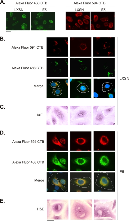
Fig. 4.
E5 promotes perinuclear membrane fusion. (A) HECs that stably express HPV-16 E6 were infected with a retrovirus encoding E5 (or harboring the empty pLXSN expression vector). Three days later, the cells were labeled for 30 min at 4°C with Alexa Fluor 488 (green) or Alexa Fluor 594 (red) conjugates of CTB, washed, and fixed. Similar levels of CTB bind to the plasma membrane of E5- and LXSN-infected cells. (B) E6-HECs were infected with a retrovirus containing the empty pLXSN expression vector and, 3 days later, were pulse-labeled for 30 min (at 37°C) with Alexa Fluor 594 CTB and Alexa Fluor 488 CTB 6 h apart. Small, nonmerged vesicles labeled with Alexa Fluor 594 CTB (red) and Alexa Fluor 488 CTB (green) were present in the perinuclear area in ca. 95% of the cells (merge). Nuclei (blue) and the boundaries of cells (white lines) are indicated. (C) H&E staining of LXSN/E6-HECs reveals morphologically similar nonvacuolated cells. (D) E6-HECs were infected with a retrovirus encoding E5 and pulse-labeled as in panel B. Large perinuclear areas of complete Alexa Fluor 488 CTB and Alexa Fluor 594 CTB colocalization were present in ca. 15% of the cells (merge). (E) H&E staining of E5/E6-HECs shows morphologically similar vacuolated cells. Scale bar, 10 μm.

