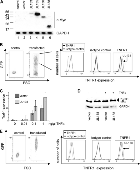
Fig. 6.
UL138 upregulates TNFR1 surface expression in HeLa and THP-1 cells and potentiates TNF-α-induced NF-κB signaling. HeLa cells were transfected with lentiviral vectors expressing either the UL133, UL135, UL136, or UL138 gene in frame with a c-Myc epitope tag and, in addition, GFP. (A) Viral protein expression was detected by immunoblotting using a c-Myc-specific antibody. Equal protein loading was controlled by including GAPDH. (B) High-GFP-expressing HeLa cells were gated for each of the UL133-UL138-transfected populations as shown (boxed), and gated cells were analyzed for TNFR1 surface expression as described for Fig. 3D. Expression analyses are shown as overlay histograms. (C) Mock- and UL138-transfected HeLa cells were puromycin selected and stimulated with TNF-α for 2 h, employing the indicated concentrations. RNA was prepared, and Traf-1 expression was analyzed by real time-PCR with normalization to GAPDH expression. (D) Transfected cells as described for panel C were treated with the proteasome inhibitor ALLN for 1 h, and phosphorylation of IκBα was detected by immunoblotting in the absence or presence of 0.01 ng/ml TNF-α (10 min). Equal protein loading was controlled for by including GAPDH. (E) TH-P1 cells were either left untransduced or were transduced with an empty lentivirus or a UL138-expressing lentivirus and puromycin selected as indicated by GFP detection. TNR1 surface expression was analyzed in all three populations, and TNFR1 surface expression is shown in the overlay histograms.

