Fig. 7.
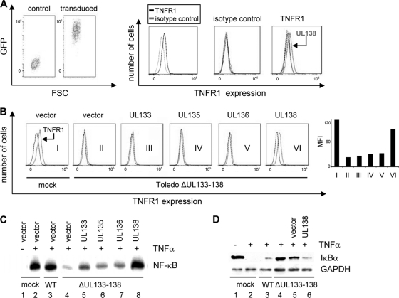
UL138 rescues TNFR1 surface expression and TNF-α-induced NF-κB activation in ToledoΔUL133-138-infected cells. (A) Primary fibroblasts were transduced with lentiviruses expressing UL133, UL135, UL136, or UL138 and puromycin selected to yield a population of greater than 99% transduced cells as indicated by GFP detection. TNR1 surface expression was analyzed in each of the GFP-positive populations expressing one of the viral UL133 to UL138 genes as described for Fig. 6. (B) Primary fibroblasts transduced with lentiviral vectors for UL133 to UL138 were either mock infected or infected with the ToledoΔUL133-138 mutant virus as indicated. At 72 h post-HCMV infection, surface expression of TNFR1 was analyzed (graphs I to VI). The bar chart shows a quantitative analysis (mean fluorescence intensity minus that of an isotype control) of the depicted histograms generated by using the FlowJo analysis software (Becton Dickinson). (C) Primary fibroblasts transduced with either control lentivirus or the indicated UL133- to UL138-expressing lentiviral constructs were superinfected with Toledo wild type (WT; lane 3), ToledoΔUL133-138 (lanes 4 to 8), or mock infected (lanes 1 and 2). After TNF-α stimulation for 10 min, cells were harvested, nuclear extracts were prepared, and NF-κB DNA binding was detected by EMSA. (D) Control cells (lanes 1 and 2), Toledo WT-infected cells (lane 3), and ToldeoΔUL133-138-infected cells (lanes 3 to 6) either transduced with a lentiviral vector (lane 5) or a UL138-expressing lentivirus (lane 6) were stimualted with TNF-α for 10 min (lanes 2 to 6). The abundance of IκBα was detected by immunoblotting. Equal protein loading was controlled for by including GAPDH.

