
Fig. 9.
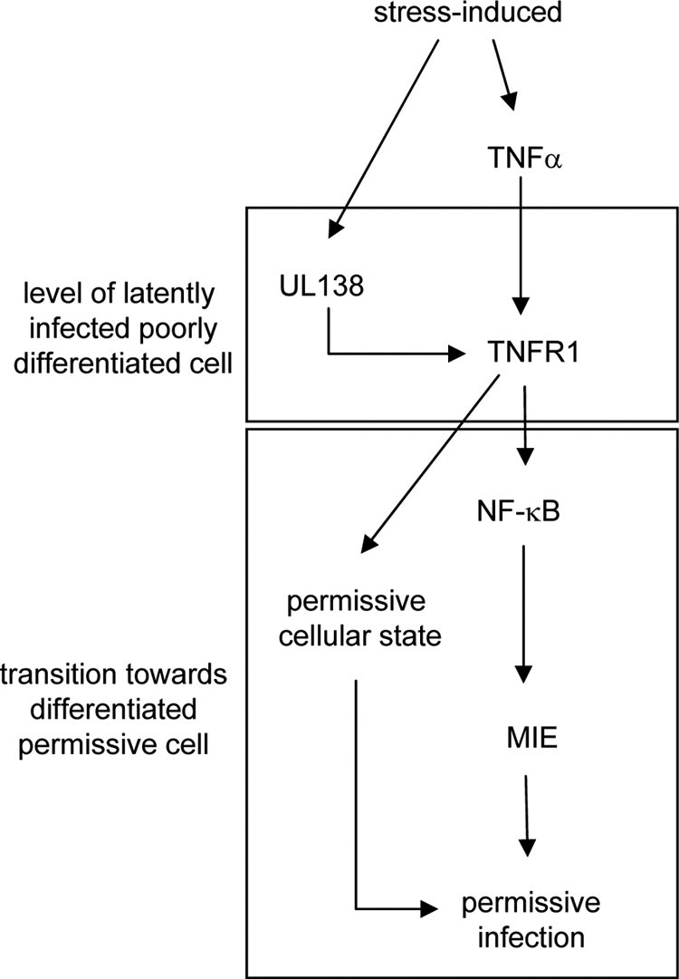
Hypothetical schematic concerning the UL138-TNFR1 interplay. For this hypothetical setting, a latently infected, poorly differentiated cell was considered. However, it is neither suggested that the UL138-TNFR1 interaction is sufficient to drive reactivation, nor has it been shown that this functional interaction exists in latently infected cells. Rather, this model serves as a rationale to envisage a biological function of this interaction with respect to published evidence. Since during latency only a very limited number of viral genes are expressed, those viral activities downregulating TNFR1 expression during lytic infection are likely to be absent at this stage and, therefore, were omitted from this model. Under stress conditions, UL138 becomes upregulated (17) and induces TNFR1 surface expression that allows TNF-α-mediated signaling (present study). TNF-α signaling is conducive to the development of an HCMV-permissive state in differentiating cells (41) and promotes, via NF-κB, viral major IE gene expression (6). Together, this contributes to a cellular and viral state that could foster reactivation.