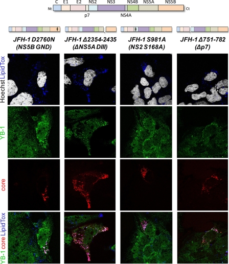
Fig. 8.
YB-1 localization phenotype of various replication- and assembly-defective JFH-1 mutants. A schematic representation of each JFH-1 mutant is shown above the corresponding panel. JFH-1 mutant-expressing Huh7.5 cells were fixed and probed with rabbit anti-YB-1 (green) and mouse anti-core (red) antibodies. Nuclei and lipid droplets were stained with Hoechst (white) and LipidTox (blue) dyes, respectively. Merged images are shown.

