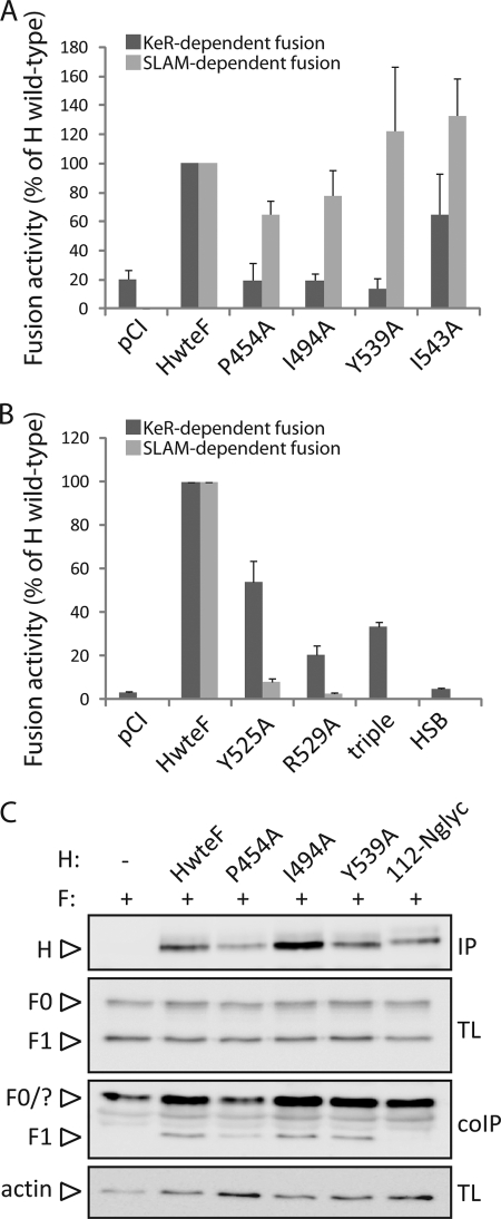
Fig. 4.
Quantitative investigation of the selectivity of receptor-dependent fusion support deficiency for selected H mutants. (A) Assessment of KeR-dependent fusion-defective H mutants in a luciferase reporter gene content-mixing assay. Vero-SLAM cells or keratinocytes (target cells) were infected with MVA-T7 (MOI, 1). In parallel, a second population (Vero cells, used as effector cells) was transfected with the different H proteins, a plasmid encoding Fwt, and a plasmid containing the luciferase reporter gene under the control of the T7 promoter. Twelve hours after transfection, effector cells were mixed with target cells (either with Vero-SLAM cells or with keratinocytes) and were seeded into fresh plates. After 2.5 h (Vero-SLAM cells) or 6 h (keratinocytes) at 37°C, fusion was quantified by using a commercial luciferase-measuring kit. For each experiment, the value for the Fwt/Hwt combination was set to 100%. Means and standard deviations for three independent experiments carried out in duplicate are shown. (B) The experiment described for panel A was carried out with previously identified SLAM-dependent fusion-defective H mutants. (C) Assessment of interaction of H with functional F proteins. To stabilize the F-H interactions, transfected Vero cells were treated, or not, with the membrane-permeant cross-linker DSP. Then Vero cells cotransfected with the various H-expressing plasmids or an empty plasmid (pCI) together with Fwt-expressing plasmids or pCI were lysed with the stringent RIPA buffer, followed by immunoprecipitation (IP) of H with a linear-epitope-recognizing anti-H MAb (1347) and treatment with protein G-Sepharose beads. Proteins were then boiled and subjected to immunoblotting using a polyclonal anti-HA antibody to detect the F antigenic materials (F contains an HA tag fused at its C-terminal region [24]). Co-IP F proteins were detected by comparison with F present in the lysates prior to IP by immunoblotting using a polyclonal anti-F antibody (TL). As a control, total H proteins, obtained by direct immunoprecipitation with the MAb mentioned above, were revealed by immunoblotting using a polyclonal anti-H antibody (IP). Finally, as a loading control, total expression of the actin protein in each sample is shown (actin).

