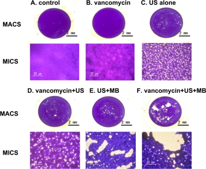
Fig. 3.
Morphology of biofilms examined by macroscopy (MACS) and light microscopy (MICS) (original magnification, ×200). Overnight cultures of bacteria were diluted 1:200 and cultured in 96-well plates (200 μl/well) at 37°C for 12 h, and then the biofilms underwent the six different treatments outlined in the figure (A to F). The biofilm was then washed gently 3 times with PBS and stained with crystal violet. After treatment, the biofilms were stained with 2% crystal violet.

