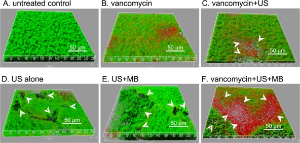
Fig. 6.
(A to F) Three-dimensional structural images of biofilms. The biofilms of S. epidermidis RP62A in FluoroDishes were visualized by CLSM with the LIVE/DEAD viability stain (SYTO9/PI). Viable cells exhibit green fluorescence, whereas dead cells exhibit red fluorescence. Many micropores (white arrow) could be observed in the biofilms treated with US with or without MB. The images are three-dimensional reconstructions using Imaris software, based on CLSM data at approximately 0.5-μm-depth increments. The experiments were repeated twice with similar results.

