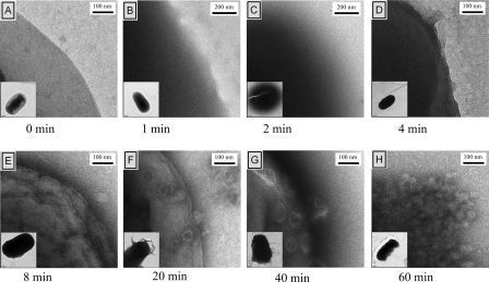
Fig. 3.
Changes in cell morphology upon treatment with PMX 10072. Cultures of E. coli D31 were incubated with 62.5 μg/ml (10× MIC) of PMX 10072 for the indicated times, stained with uranyl acetate, and visualized by TEM. (A) The membrane is distinct and uniform prior to treatment. (B) After 1 min exposure, the cytoplasm appears dark due to increased stain accessibility. (C) A diffuse halo appears around the cell 2 min after exposure. (D) By 4 min after treatment, the cells reestablish cellular integrity, although they continue to be permeable to stain. (E) The membrane becomes more nonuniform with extensive ruffling with exposure for 8 min. (F) Vesiculation of the outer membrane occurs after 20 min of exposure. (G and H) Vesiculation continues to worsen (G) and results in the total loss of membrane integrity (H).

