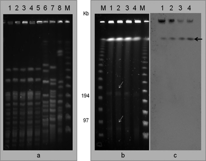
Fig. 1.
(a) PFGE analysis of Acinetobacter baumannii strains. (b) Plasmid identification by digestion with S1 nuclease. (c) Hybridization with blaNDM-1 probe. Lanes: 1, A. baumannii AB-I1; 2, AB-I2; 3, AB-I3; 4, AB-I4; 5, AB-I5. Lanes 6 to 8, A. baumannii European clones EC-I (strain RUH-875), EC-II (strain RUH-134), and EC-III (strain RUH-5875), respectively. Bands with white arrows indicate the presence of plasmids without signal hybridization with the blaNDM-1 probe; black arrow indicates the chromosomal position with positive hybridization with the blaNDM-1 probe.

