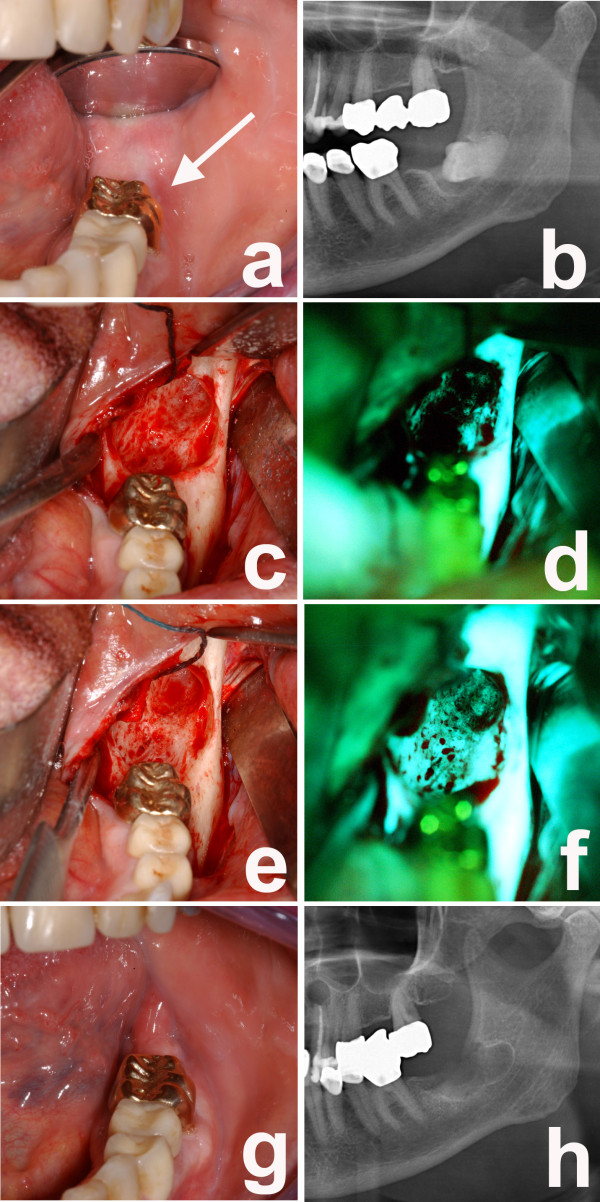
Figure 1.
a) Intraoral examination of her left upper jaw with fistula formation and pus on palpation in region 23; b) dental X-ray examination with gutta-percha pin in the fistula; c) intraoperative view with bony defect in region 23; d) fluorescence optic view with loss of fluorescence in region; e) bone cylinder region 23 with f) a mild fluorescence in the superficial areas and almost complete loss of fluorescence in deeper areas; g) corresponding clinical picture of the bone cylinder; h) histological examination of a representative biopsy with necrotic bone.

