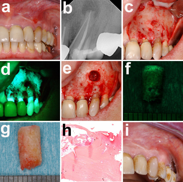
Figure 2.
a) intraoral examination of her left lower jaw with fistula formation and pus on palpation in region 38; b) panoramic radiograph with mixed radiopaque and radiolucent areas surrounding the retained wisdom tooth 38; c) intraoperative situs after wisdom tooth removal; d) corresponding fluorescence picture with loss of fluorescence in the lingual aspects of region 38; e) intraoperative situs after removal of necrotic bone parts; f) corresponding fluorescence picture with markedly enhanced fluorescence in the lingual aspects of region 38; g) intraoral examination eight weeks postoperatively with complete mucosal closure and without fistula formation; h) panoramic radiograph after removal of the wisdom tooth 38 and necrotic bone parts; i) intraoral examination eight weeks postoperatively with complete mucosal closure and without fistula formation.

