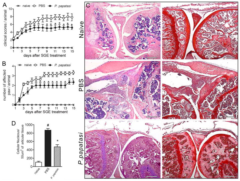
Figure 1. SGE from P. papatasi attenuated collagen-induced arthritis.
Naïve (×) or collagen-immunized and challenged DBA/1 mice were injected i.v. daily with PBS (○) or P. papatasi SGE (1 gland/animal) (●) for 14 d. Mice were monitored for disease progression as indicated by clinical scores (A)and number of affected paws(B). On d15 of SGE treatment, mice were euthanized, the articular joints were harvested, and histopathologic analysis was performed. Knee joint sections were stained by H&E (left row) or with safranin-O (right row), a proteoglycan red marker showing profound cartilage damage in PBS-treated mice (absence of proteoglycan) and preservation of cartilage in SGE-treated mice(C). Quantification of cellular infiltrate was performed by ImageJ software (NIH, USA)in 40 fields with 400× magnification for each animal/group (D). Morphometric histologic examination shows markedly less cellular infiltration in the SGE-treated than in the PBS-treated group (black bar vs. striped bar). Results show the mean±SEM, N =4; *, P<0.05 compared with PBS-treated group.

